Non-fluorescence techniques
At the light microscopy part of CCI the focus is on advanced fluorescence techniques. But on all of our microscopes the fluorescence imaging can be combined with other contrast techniques, such as brightfield, phase contrast or DIC. On some of our microscopes we can also set up polarization or darkfield, when needed.
Brightfield imaging is the simplest light microscopy technique, where usually white light is either transmitted through (biological science) or reflected by (material science) the sample. Köhler illumination settings make sure that there is an uniform illumination intensity at the sample plane.
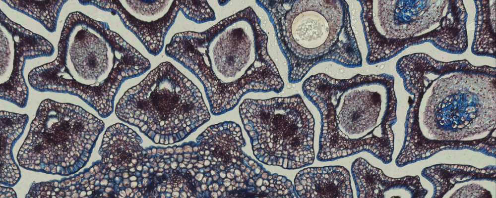
In a widefield microscope, equipped with a color camera, it is possible to visualize a biological sample labeled with histological stains, e.g. Hematoxylin and Eosin. At the CCI there are color cameras at the AxioObserver microscope and at the PALM Microbeam LMD system.
On all confocal microscope systems there is a detector in the transmission beam path, in addition to the detectors for fluorescence detection in the reflection beam path, which allows for brightfield imaging using any of the lasers.
In darkfield microscopy the central parts of the illumination is blocked, and only light passing through the specimen from oblique angles is diffracted, refracted, and reflected into the microscope objective to form a bright image of the specimen on a dark background.
This dark background provides a high degree of contrast and can make small samples or samples with difficult backgrounds visible.
There is currently a darkfield condenser (dry darkfield condenser 0.8/0.95 (0.6-0.75) WD=6.0mm) which can be installed on for example the AxioObserver microscope.

