
Algorithm helps doctors identify more aggressive types of basal cell carcinoma
An algorithm can help healthcare professionals recognize which patients have a highly aggressive form of basal cell carcinoma (BCC) of the face. These are the findings of a study conducted at the University of Gothenburg. If more BCCs are correctly identified as high-risk, the patients can directly receive the most effective treatment.
BCC is the most common form of skin cancer. The cancer type grows slowly and almost never spreads to other parts of the body. Most of the BCCs all are cured, but without treatment, highly aggressive forms can grow infiltratively and cause significant morbidity for affected patients. It is a growing cancer type, with 70,000 new cases confirmed in Sweden in 2021.
The most obvious signs
The study includes clinical as well as dermoscopic images obtained at the Department of Dermatology and Venereology at Sahlgrenska University Hospital. Images of nearly 300 patients with confirmed facial BCC were analyzed. These images were then reviewed by six independent experienced dermatologists who were asked to interpret the observed clinical and dermoscopic findings in each case. These findings were subsequently used as the basis for the development of a clinical algorithm that serves to discriminate between low and more aggressive types of BCC.
The study pinpointed that the BCCs with a bumpy surface had a strong association with high-risk subtype of BCC. Moreover, poorly defined borders, and presence of a lighter area (often called “white porcelain area”) were also associated with this high-risk subtype. Small blood vessels in the ulceration were also found to be characteristic of an aggressive form, which was not previously known.
The study shows that the clinical algorithm identifies most cases of high-risk BCC. The algorithm also showed a high positive predictive value, i.e., when it flags a BCC as high-risk, it is usually right.
The best treatment
![[none]](/sites/default/files/styles/33_10_13_small_1x/public/2024-06/Hannah%20Ceder%20foto%20Paul%20Bj%C3%B6rkman.jpg?h=5dde4fee&itok=8lVJriJ8)
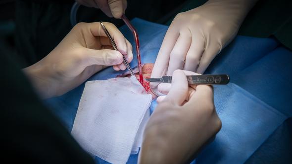
The first author of the study, Hannah Ceder, is a PhD student at the University of Gothenburg and a specialist physician at the Department of Dermatology and Venereology at Sahlgrenska University Hospital. She is also one of the surgeons in the country who performs the surgical method that has been shown to be clearly superior for high-risk BCCs. The surgical method is called Mohs micrographic surgery, which allows for complete examination of all tissue margins guaranteeing complete removal, while sparing as much healthy tissue as possible. While the patient is under local anesthesia on the operating table, pathologists analyze the tissue samples to be absolutely sure that the entire tumor is gone, before closing the surgical wound.
“Mohs micrographic surgery gives us full control of the margins, while preserving healthy tissue. We have shown in a previous study that in patients with highly aggressive BCC of the nose, traditional surgery fails to remove the entire tumor in over half of the cases,” says Hannah Ceder.
Distinguishing the tumors
In other surgeries, there is a risk that the surgery will have to be repeated because the pathologist will find residual tumor in the tumor margin. However, waiting times for Mohs micrographic surgery are long: in Gothenburg currently about one year.
“It is desirable to develop simple preoperative methods that help doctors in identifying these high-risk tumors. Therefore, our clinical algorithm is relevant and important. This would make it easier to determine which tumors can be easily removed with traditional surgery without a prior biopsy, and which ones require further investigation to screen out the cases that require Mohs micrographic surgery,” says Hannah Ceder. However, it is important to revalidate the clinical algorithm in a prospective setting to investigate its usefulness in a real-world clinical setting.