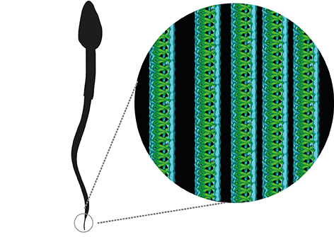
A highly effective tail is needed in order for a sperm to be able to swim, and for a baby to be conceived. By using cryo-electron tomography, researchers at the University of Gothenburg – working in partnership with researchers in the USA – have identified a completely new nanostructure inside sperm tails.
Human sperms are incredibly important for our reproduction. It would therefore be easy to assume that we have detailed knowledge of their appearance. However, an international team of researchers has now identified  a completely new nanostructure inside sperm tails, thanks to the use of cryo-electron tomography.
The method, for which Joachim Frank, Jacques Dubochet and Richard Henderson were awarded a Nobel Prize in 2017, produces 3D images of cellular structures.
“Since the cells are depicted frozen in ice, without the addition of chemicals which can obscure the smallest cell structures, even individual proteins inside the cell can be observed” explains Johanna Höög, a research at the University of Gothenburg’s Department of Chemistry and Molecular Biology.
A highly effective tail is needed in order for a sperm to be able to swim, and for an egg to be fertilised.
 The tail is a highly complex machine that consists of around a thousand different types of building blocks. The most important of these are called tubulins, which form long tubes (microtubules). The tubes are found inside the sperm tail.
Thousands of motorproteins – molecules that can move – are affixed to these tubes. By being fixed to one microtubule and “walking on” the adjacent microtubule, the motorproteins in the sperm tail pull and the tail bends, enabling the sperm to swim.
“It’s actually quite incredible that it can work,” adds Johanna, who led the study. “The movement of thousands of motorproteins has to be coordinated in the minutest of detail in order for the sperm to be able to swim.”
The research was started in order to see what human sperm tails look like in 3D. This would then provide clues about how sperms work, in the same way that a sketch of an engine helps to explain how it operates.
“When we looked at the first 3D images of the very end section of a sperm tail, we spotted something we had never seen before inside the microtubules: spiral that stretched in from the tip of the sperm and was about a tenth of the length of the tail.”
What the spiral is doing there, what it consists of and whether it is important  in order for sperms to swim are questions that the research team will now focus on answering.
“We believe that this spiral may act as a cork inside the microtubules, preventing them from growing and shrinking as they would normally do, and instead allowing the sperm’s energy to be fully focussed on swimming quickly towards the egg,” says Davide Zabeo, the lead PhD student behind the discovery.
The study, which has now been published in the journal Scientific Reports, is a collaboration between researchers at the University of Gothenburg in Sweden and the University of Colorado in the USA.
Title of the article: A lumenal interrupted helix in human sperm tail microtubules.
Link to article>>
Contact details:
Johanna Höög, researcher at the Department of Chemistry and Molecular Biology, University of Gothenburg, +46 (0)31 786 39 37, +46 (0)766 18 39 37, johanna.hoog@gu.se