Events at CCI
The CCI regularly organizes conferences, workshops and seminars in collaboration with other universities in Sweden and abroad and with industrial partners
CALL4HELP: Monthly event
CALL4HELP in Light Microscopy, Image Acquisition & Analysis
Are you struggling with your imaging pipeline and need advice on which microscopy technique is best for your sample, what analysis workflow would best answer your question or simply how to set up the whole imaging experiment?
CCI can help!
On the 3rd Friday of each month we are organising open hours (13:00 – 15:00) where our light microscopy and bioimage analysis experts can meet with you and discuss your sample preparation protocol (if you share it in advance), which microscope to use, which microscopy modality to use (e.g. functional microscopy), how to generate appropriate controls, how to perform quantitative image analysis, and other topics.
To register follow the link to the Registration Form
Registration:
Where do we meet?
We meet in the computer room located at the light microscopy corridor of the CCI. Medicinaregatan floor 1, next to the elevator close to entrance 1F
CAT workshop 2023
From November 29th to December 1st, 2023, the Centre for Cellular Imaging hosted the Cellular 3D Volume – Correlative Array Tomography (CAT) Workshop. This workshop introduced participants to the cutting-edge technology of array tomography, a powerful technique combining light and electron microscopy for exploring three-dimensional cellular architectures of large samples in fine molecular and structural detail.
The event brought together experts, researchers, and industry professionals to share knowledge and foster collaborations through a mix of lectures and hands-on demonstrations. Topics covered included volume electron/light microscopy, sample preparation for array tomography, image processing and analysis, correlative techniques, and multibeam scanning electron microscopy. The workshop provided an excellent opportunity to network, exchange ideas, and learn new methods for advancing research into complex 3D tissue architectures.
Workshop website
Opening of the Lightsheet 7
Practical information
Event Timing: January 18th, 2024
Lectures Address: Birgit Thilander, Academicum, Medicinaregatan 3
Demo Address: Medicinaregatan, floor 1, staircase of door 7A

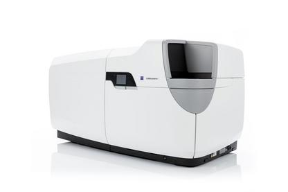
The Centre for Cellular Imaging is pleased to announce the purchase and installation of the Lightsheet 7 fluorescence microscope. Light sheet fluorescence microscopy (LSFM) uses a thin sheet of light to excite only fluorophores within the focal volume. It is ideal for fast and gentle imaging of whole living model organisms, tissues and cells as they develop – over extended periods of time. Furthermore, the Lightsheet 7 is designed to handle large optically cleared specimens with subcellular resolution. Dedicated optics, sample chambers and holders allow adaption to the refractive index of the chosen clearing method.
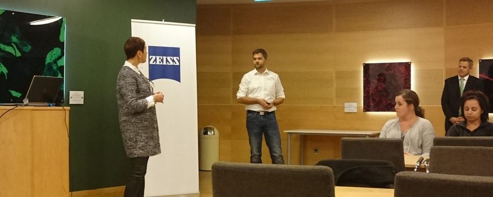
The Lightsheet 7 is now installed at the Centre for Cellular Imaging and to celebrate this we will host an "Opening Day" with seminars and demos, where you can learn about this technology and other types of equipment available at the CCI.
Schedule
| Time | Event |
|---|---|
| 9:00-9:10 | Welcome from the Head of the Centre for Cellular Imaging, Dr Julia Fernandez-Rodriguez. |
| 9:10-10:10 | Lightsheet 7 - Dr. Jacques Paysan, Product & Application Sales Specialist, ZEISS |
| 10:10-11:10 | The Arivis Scientific Imaging Platform, Molly McQuilken, Business Development Manager, ZEISS |
| 11:10-13:00 | Lunch break |
| 13:00-15:00 | Visit of the Centre for Cellular Imaging and demos |
Opening day at the Centre for Cellular Imaging Elyra 7 and LSM980
LSM980 with Airyscan 2 and Elyra 7 with Lattice SIM and SMLM
The Centre for Cellular Imaging (CCI) is pleased to announce the purchase and installation of two new systems: A Super-resolution and a Laser Scanning Microscope for fast live imaging!!
| Event Timing: | February 23rd, 2023 |
| Lectures Address: | Hörsal Arvid Carlsson, Academicum, Medicinaregatan 3 |
| Demo Address: | Medicinaregatan, floor 1, staircase of door 7A |
ZEISS ELYRA 7 with Lattice SIM² and Single Molecule Localization - Your Live Imaging System with Unprecedented Resolution. The super-resolution microscope ELYRA 7 with Lattice SIM² you can now double the conventional SIM resolution and discriminate the finest sub-organelle structures, even those no more than 60 nm apart. You don‘t need to sacrifice resolution when imaging at high speed (up to 255 fps ) using only the minimal exposure needed for life observation. ELYRA 7 enables you to combine super-resolution and high-dynamic imaging – without the need for special sample preparation or expert knowledge of complex microscopy techniques.
ZEISS LSM 980 with Airyscan 2 - Your Unique Confocal Experience for Fast and Gentle Multiplex Imaging. To analyze life with as little disturbance as possible, you must use low labeling density for your biological models. This requires excellent imaging performance combined with low phototoxicity and high speed. LSM 980, your platform for confocal 4D imaging, is optimized for simultaneous spectral detection of multiple weak labels with the highest light efficiency.
Venue for the lectures: Hörsal Arvid Carlsson, Academicum, Medicinaregatan 3 Google Maps link
Schedule 23rd of Feb 2023
| Time | Event |
|---|---|
| 9:50 - 10:00 | Welcome from the Head of the Centre for Cellular Imaging, Dr Julia Fernandez-Rodriguez |
| 10:00 - 10:35 | LSM 980 with Airyscan 2 – Your Unique Confocal Experience for Fast and Gentle Multiplex Imaging. Dr Chris Power, Product & Application Sales Specialist, ZEISS Research Microscopy Solutions |
| 10:35 - 11:10 | ELYRA 7 – Ways to improve decoding algorithms in Structured Illumination and Localization Microscopy. Dr Klaus Weisshart, Product Management, ZEISS Research Microscopy Solution |
| 11:10 - 11:40 | The Arivis Scientific Imaging Platform: Analysis without Compromises, Molly McQuilken, Business Development Manager, Arivis at ZEISS company |
| 12:00 - 13:00 | Lunch Break |
| 13:00 - 15:00 | Visit to the CCI Facility and demos |
Open lectures, 18th of May - 21th of May, 2021: Artificial intelligence and bio-image analysis
As part of the "Smart Microscopy" workshop, see above, we have invited external speakers to give lectures on relevant topics in artificial intelligence and bio-image analysis (Deep Learning, Tissue analysis, Quantitative microscopy, Big Data handling, etc.). On this occasion, we are very fortunate to welcome:Carolina Wählby (SciLifeLab - Uppsala University, Sweden), David Dang (London's Global University - UK) and Jean-Yves Tinevez (Institut Pasteur, France). These lectures are open to anybody interested in these topics but registration is mandatory.
- Monday 17th of May, 13:00-13:30 CEST: Aliaksandr Halavatyi, "Designing adaptive feedback microscopy pipelines and performing experiments with AutoMicTools library in Fiji"
Lecture: https://youtu.be/7w9khqssZrw - Tuesday 18th of May, 13:00-13:30 CEST: David Dang, "SpinX: Time-resolved 3D Analysis of Spindle Dynamics using Deep Learning Techniques and Mathematical Modeling"
Lecture: https://youtu.be/7_SwQ4Cd1ME - Thursday 20th of May, 13:00-13:30 CEST: Carolina Wählby, "Image analysis and AI in microscopy-based life science research"
Lecture: https://youtu.be/EdiX1tJUSxk - Friday 21st of May, 13:00-13:30 CEST: Jean-Yves Tinevez, "Tracking cells for automated microscopy"
Lecture: https://youtu.be/mLUeSkpI07w
ONLINE opening day: Celldiscoverer 7 / LSM 900 Airyscan 2, 30 March, 2021
The Centre for Cellular Imaging is pleased to announce the purchase of an automated High Content Screening system combined with a Laser Scanning Confocal Microscope: the Celldiscoverer 7/ LSM 900 Airyscan 2 from Zeiss. With this system we open up the possibility for advanced automated live cell imaging with feedback experiments and guided acquisition including advanced image processing and analyzing capability! For more information, visit the Zeiss web page
This microscope is now installed at the CCI and to celebrate this, we will host an “ONLINE opening day” with seminars where you can learn about the advanced technology that powers this class-leading instrument. During this day we would like to invite you to take a glimpse of what is possible with today’s advanced automated live cell imaging system, and what type of questions can be addressed.
Registration:
Register to juliafer@cci.sahlgrenska.gu.se by March 26th. On March 29th you will receive the link for the Seminar
Program of the day:
13:00 - 13:15 Welcome and introduction; Dr Julia Fernandez Rodriguez, Head of the Centre for Cellular Imaging, and Dr Jens Wigenius, Zeiss Regional Sales Manager
13:15 - 13:45 "Advanced automated Live cell imaging with Zeiss Celldiscoverer 7/LSM 900 Airyscan2”; Dr. Sören Prag, Zeiss Application specialist in automated and advanced imaging systems
13:45 - 14:00 Short-break
14:00 - 14:20 Life Sciences Applications; Dr. Sören Prag, Zeiss Application specialist in automated and advanced imaging systems
14:20 - 15:00 Q&A
15:00 - 16:00 Live virtual demonstration Zeiss Celldiscoverer 7/LSM 900 Airyscan2; Dr Rafael Camacho, Scientific Officer Centre for Cellular Imaging, and Dr Maria Trulsson, Zeiss Application specialist

